Disclaimer: This article is intended solely for informational and educational purposes only. It does not constitute medical advice.
Regional anesthesia has transformed pain management by offering increasingly precise and targeted methods for controlling pain while also reducing exposure to systemic anesthetic drugs. Ultrasound techniques have increased this precision, since these procedures allow clinicians to visualize nerves and their surrounding structures in real time. However, even with the advent of advanced imaging systems, anatomical variations in nerves, blood vessels, and nerve roots can prove to be obstacles in otherwise routine regional anesthesia procedures. Differences in where nerves originate, where they branch, and their pathways can affect whether an anesthetic drug reaches its target and may introduce complications such as puncture injury or other related risks.(1) Understanding these variations is essential to making regional anesthesia safer, more predictable, and more effective.(2)
The terminal branches of the trigeminal nerve include the supraorbital nerve, infraorbital nerve, and mental nerve; these three divisions provide sensation to different areas of the face. All three nerves show considerable variation around their exit foramina, including accessory foramina and differences in branching patterns. For example, the supraorbital notch/foramen is located approximately 26 mm from the midline in Caucasian populations, while the difference is only about 22 mm in Asian populations.(3) Incomplete block could also arise due to the infraorbital nerve branching prematurely within the infraorbital canal and exiting through an accessory foramen, which can be found in up to 38.7% of Chinese individuals.(4)
Additionally, the mental nerve can have an anterior loop or multiple exit points; this varies significantly with ethnicity. These variations are all potential causes of incomplete anesthesia if the local anesthetic is deposited only around the expected main nerve exit. Anesthesia may not spread to accessory foramen, as these require a wider or additional injection. Altered nerve courses can also increase the risk of neurovascular injury.(5)
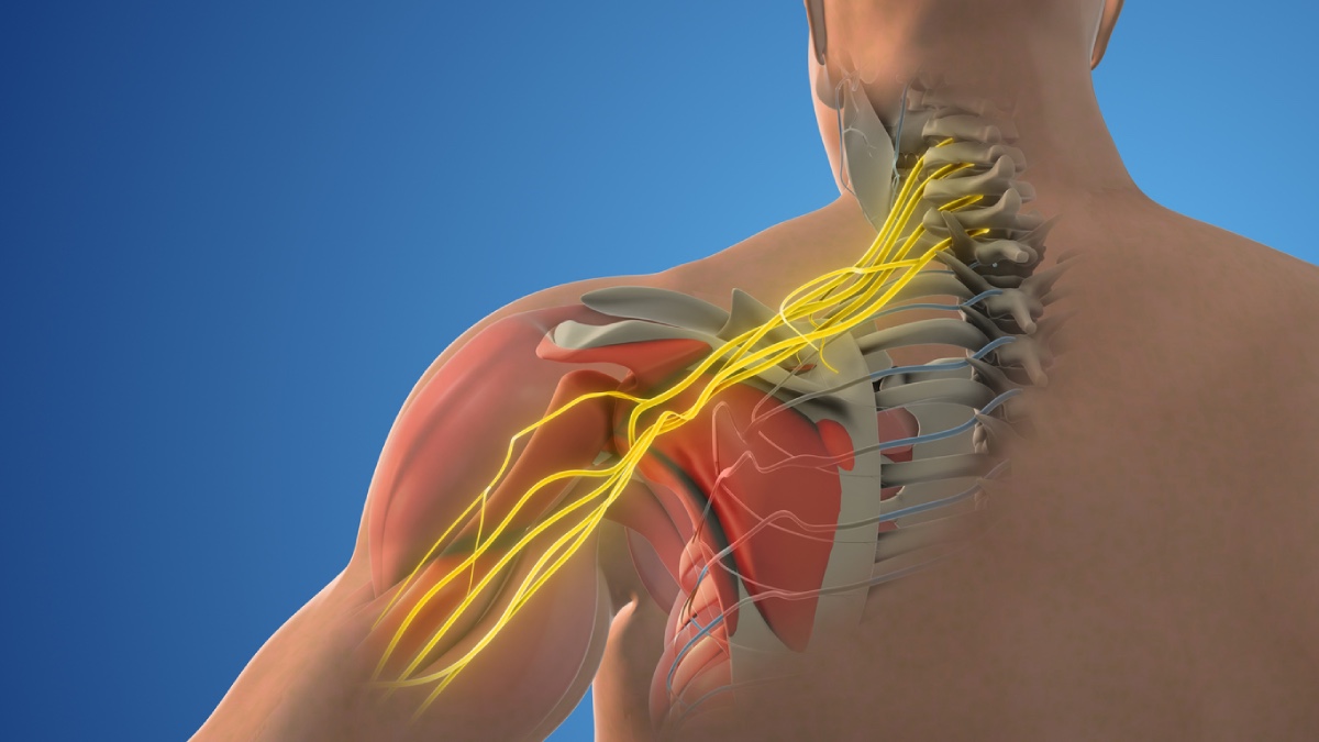
The phrenic nerve is the major motor nerve of the diaphragm; blocking this nerve can impair diaphragmatic movement. The effect of anatomical phrenic nerve variations on regional anesthesia is particularly important during interscalene and supraclavicular brachial plexus blocks, since there is a risk the local anesthetic may spread beyond the intended target and cause diaphragmatic impairment or paralysis. Individuals with variations that bring the phrenic nerve closer to the brachial plexus increase the risk of this specific complication.(2)
The lumbar plexus is a network of nerves that lies deep within the psoas major muscle and gives rise to the femoral, obturator, and lateral femoral cutaneous nerves. The femoral nerve is especially important for regional anesthesia because it provides motor and sensory innervation to most of the anterior thigh and the medial leg.(6) Because a lumbar plexus block relies on the drug effectively spreading through the psoas department to reach multiple nerves, anatomical variations in the origins, relationships, and courses of lumbar plexus branches can affect which nerves are successfully blocked and how reliable that block is. The deep location of the plexus also increases the need for accurate ultrasound identification and needle placement.(2)
Anatomical variations in nerves is an important consideration for regional anesthesia, especially as ultrasound becomes more widespread in clinical settings. Variations in nerve origins, branching, and pathways can make individual nerves difficult to identify, and can contribute to incomplete anesthesia, unintentional nerve block, or other related complications. Recognizing these variations allow clinicians to interpret unexpected outcomes and adapt injection sites or delivery techniques when necessary.
References
- Răzvan Costin Tudose, Mugurel Constantin Rusu, Sorin Hostiuc. The Vertebral Artery: A Systematic Review and a Meta-Analysis of the Current Literature. Diagnostics 2023;13(12):2036-2036. 10.3390/diagnostics13122036
- Zhang Y, Xu S, Lan H, Ma D, Wang Y. Clinical Implications of Anatomical Variations in Nerves and Adjacent Structures for Regional Anesthesia: A Narrative Review. Pain and Therapy 2025;15(1):41-69. 10.1007/s40122-025-00797-5
- Ashwini L, Mohandas Rao K, Saran S, Somayaji S. Morphological and Morphometric Analysis of Supraorbital Foramen and Supraorbital Notch: A Study on Dry Human Skulls. Oman Medical Journal 2012;27(2):129-133. 10.5001/omj.2012.27
- Hanihara T, Ishida H. Frequency Variations of Discrete Cranial Traits in Major Human Populations. III. Hyperostotic variations. Journal of Anatomy 2001;199(3):251-272. 10.1046/j.1469-7580.2001.19930251.x
- Kaori Katakami, Mishima A, Shiozaki K, Shimoda S, Hamada Y, Kobayashi K. Characteristics of Accessory Mental Foramina Observed on Limited Cone-beam Computed Tomography Images. Journal of Endodontics 2008;34(12):1441-1445. 10.1016/j.joen.2008.08.033
- Benes M, Zido M, Machac P, et al. Variations of the Extrapsoas Course of the Lumbar Plexus With Implications for the Lateral Transpsoas Approach to the Lumbar Spine: A Cadaveric Study. Acta Neurochirurgica 2024;166(1). 10.1007/s00701-024-06216-6